Description
General Introduction:
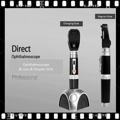
The ophthalmoscope can be divided into direct and indirect glasses.
Direct ophthalmoscope can directly check the fundus of the eye, without having to disperse the big pupil. Check in the dark room. The eye of the examiner must be close to the eyes of the patient. The right eye is used to check the right eye of the patient, the right hand is used to take the glasses, and sit on or stand on the right side of the patient. The left eye is opposite. The other hand of the doctor opens the eyelid of the patient, and first put the glasses in front of the patient about 20cm, Check the transparency of the diopter stroma with the +10d lens. After checking the diopter stroma, you can start to check the parts of the fundus. Turning the lens disc can correct the refractive errors of the doctor and the patient. If the doctor is facing the eye or the glasses are equipped with the correction glasses, you can see that the diopter used in the fundus indicates the refraction of the inspected eye.The patients were usually given a direct vision ahead, the papilla was examined, and then the temporal, infratemporal, upper and lower nasal quadrants were examined along the omental vessels. Finally, the eyes were fixed to the temporal side, and the macula was examined. The size of fundus lesions is expressed by the diameter of the optic papilla, and the concave and convex degree of the lesions are measured by the diopter of the lens. 3D is equivalent to 1mm. Some glasses are equipped with green filters, which can be used to observe optic nerve fibers and macular.
When using indirect ophthalmoscope, the pupil should be filled and dispersed. In the dark room, the doctor should turn on the power supply, adjust the distance and the position of the mirror. At first, the weak light should be used to observe the turbidity of cornea, crystal and vitreous body. Then the light should be directly injected into the pupil of the inspected eye, and the inspected eye should be fixed on the light source. Generally, the +20d objective lens should be placed 5cm in front of the eyes, The convex surface of objective lens is toward the examiner, the examiner holds the objective with his left hand and fixed it on the orbital edge of the patient. The eyes, objective lens and the head of the examinee are fixed. When the eye papilla and macula are seen, the objective lens will move towards the direction of the examiner. The stereoscopic inverted image of the optic papilla and macula can be seen clearly at 5cm in front of the examined eyes.
Products Name:
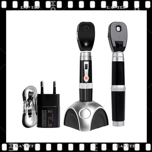
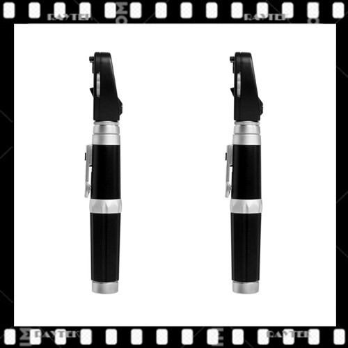
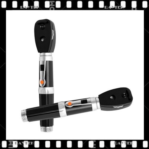
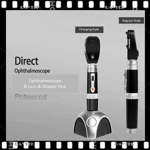
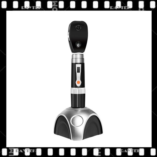
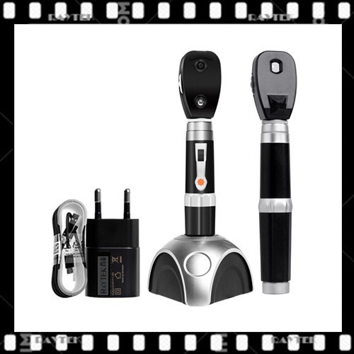
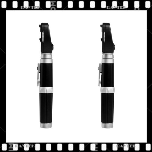
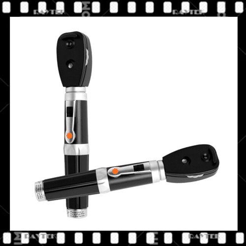
Ophthalmoscope/Direct Ophthalmoscope/Ophthalmoscope Lens/Ophthalmoscope Diopter
Disc/Ophthalmoscope Acrylic lens
Alias:Ophthalmoscope,Direct Ophthalmoscope,Ophthalmoscope Lens,Ophthalmoscope Diopter
Disc,Ophthalmoscope Acrylic lens, Acrylic lens, Optical lens for ophthalmoscope, Acrylic lens for ophthalmoscope,Ophthalmoscope Condenser, Condenser for ophthalmoscope, Condenser lens, Acrylic Condenser Lens,etc
Main Characters:
1).The coaxial vision system with enhanced optical system, high precision light source and optical system improves illumination uniformity and visibility.
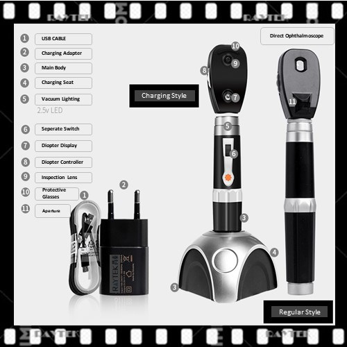
2).The diameter selection paddle provides 6 kinds of diameter(Aperture) selection:
2)-1. Microplate aperture: can quickly enter the small pupil without dilation.
2)-2. Small spot aperture: you can see the fundus of the undivided eye very well.
2)-3. Large aperture: standard aperture for mydriatic ophthalmic examination.
2)-4.Fixed aperture: graduated cross line, which can measure eccentric fixation or damage location.
2)-5. Cobalt blue filter: observe small damage, surface abrasion and corneal foreign body with fluorescent staining.
2)-6. Fissure: used to determine the level of injury and tumor
3).The lens selection wheel is simple and smooth, 68 kinds of lenses can be selected, with diopter display.
4).The high brightness HPX light source can easily penetrate cataract and other turbid bodies.
5). Medium density filter, moderate brightness into the eye and avoid reflection.
Main Technical Data: The listed prices are only for reference. The final price is up to the substrate type, substrate grade, product size, suface finish, surface quality and Physical & Optical request standards.
Main Export Countries & Areas:
Usa, Uk, Japan, Germany, Spain, France, Swiss, Korea, Russia, Pakistan, India, Portugal, Canada, New Zealand, Australia, Saudi Arab, Turkey, Finland, Poland ,etc.
Payment Method: by T/T or Western Union.
Delivery time: 7-10 days.
Quality Warranty: Ruitaiphotoelectric(Raytekoptics) offers quality warranty for our optics products with "3R" policy. For any inferior-quality products, Ruitaiphotoelectric(Raytekoptics) is responsible for return, replacement and refund.
